Spine and Brain Anatomy
It is important to know where the tumor is located within the brain and the spine so you can understand what symptoms may occur. The biology may also be different based on the location.
What Do I Need to Know About Spine and Brain Anatomy?
During treatment, your health care team will use many terms to refer to locations in the head, neck and spine. The diagrams in this section will help you locate different parts of the brain and spine. Click any of the images to see a larger version.
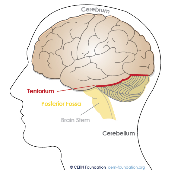
Tentorium Cerebelli
The image below shows a side (lateral) view of the tentorium cerebelli – an extension of the dura matter that separates the cerebellum from the bottom (inferior) portion of the occipital lobes. Tumors below the tentorium are called infratentorial and those above are called supratentorial.
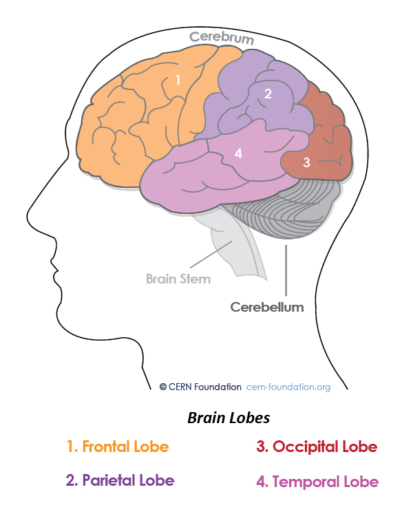
Lobes
The images show views of the lobes: side (lateral) view of the brain. The brain is divided down the middle lengthwise into two halves called the cerebral hemispheres. Each hemisphere is divided into four lobes.
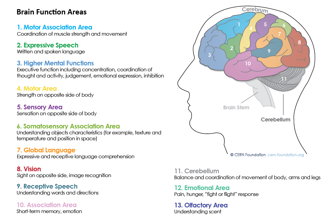
Brain Functions
See the parts of the brain to learn what they do. Knowing the location of your tumor(s) may help you to understand changes in how you act or think. Changes can be due to the impact of the tumor itself or from treatment. For example, if you have a tumor in the temporal lobe, you may have short-term memory loss. Tumors can also be associated with difficulty with multi-tasking, seizures, and headaches. It is important to address these concerns with your health care provider to evaluate if there are options to help alleviate their impact on your quality of life.
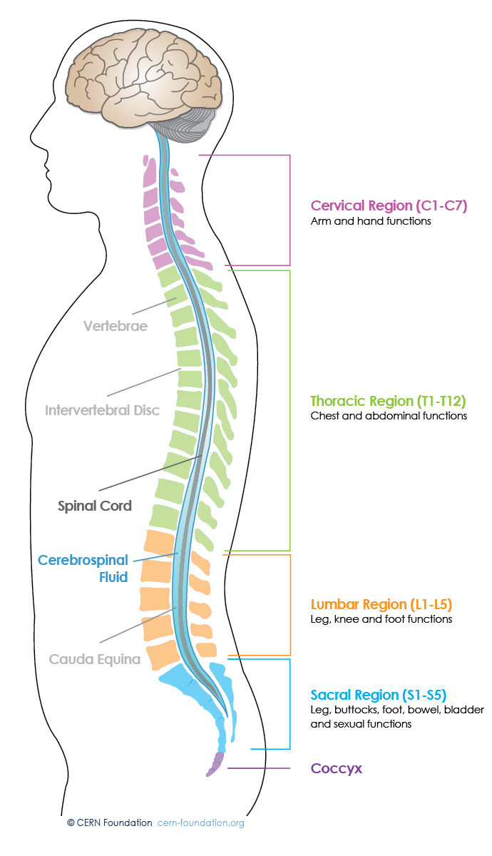
Spinal Functions
The image below shows the side (lateral) view of where the different spinal functions exist. The spine is separated into four parts called the cervical, thoracic, lumbar, and sacral region. Each spinal region is made up of vertebra. Vertebrae are numbered starting with one and continue. For example, T2 is the second vertebra in the thoracic region.
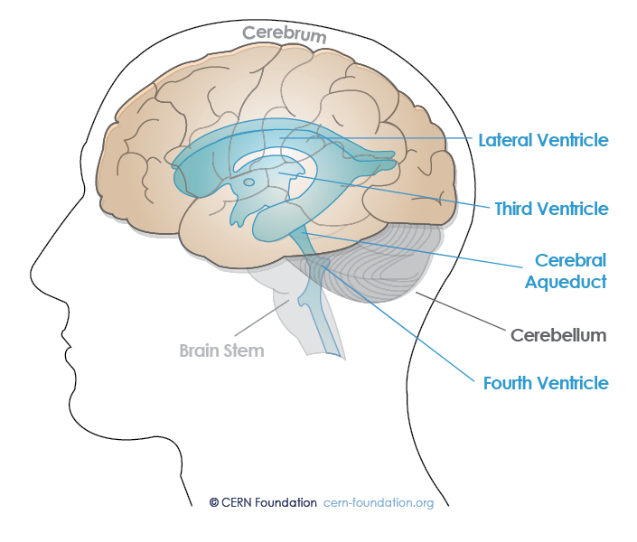
Ventricles
The image below shows the ventricular system in the brain. Ependymomas can occur anywhere in the central nervous system. Common locations include the ventricles (fluid-filled spaces in the brain that contain cerebrospinal fluid) and the central canal of the spinal cord.
Skull (Pediatric)
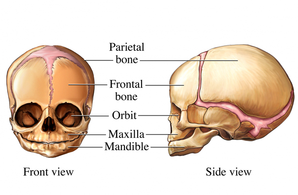
As you can see below, a child’s skull is different than an adult’s skull. Below is an image of an infant skull. When a child is born, soft spots (or fontanelles) allow the skull to shift. As the child grows, the fontanelles close up and the bones of the skull fuse.
This medical illustration details the sutures of the fetal skull. Two images show the fetal skull from a front (anterior) and side (lateral) view. Specific areas shown include the anterior fontanelle, coronal suture, frontal suture, sagittal suture and lambdoid suture.
Skull (Adult)
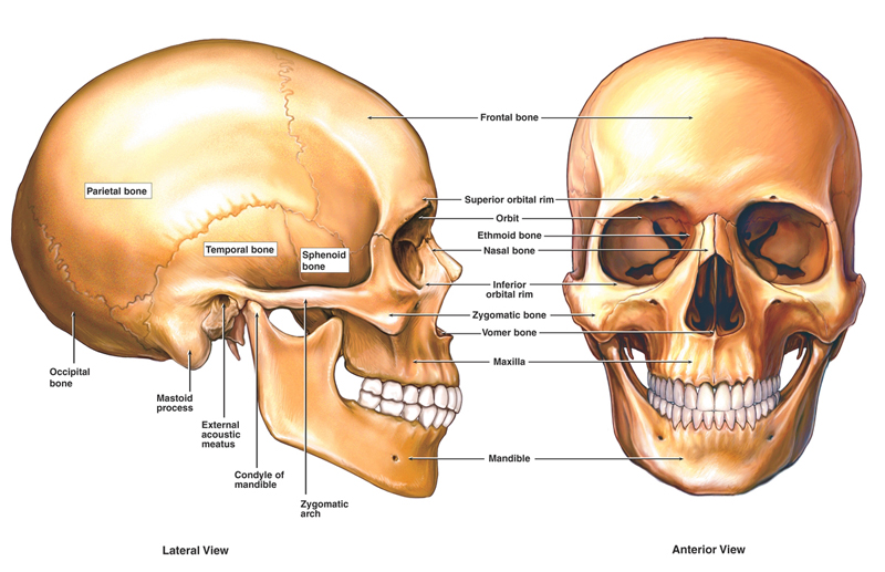
The images below accurately display the anatomy of the skull from side (lateral) and front (anterior) views. Labeled bones include the frontal bone, temporal bone, orbit, nasal bone, maxilla, mandible, zygomatic bone, zygomatic arch, mandibular condyle, external acoustic meatus, mastoid process, occipital bone and parietal bone.
For more information about brain and spine anatomy, view our Ependymoma Guide.
Support the work of CERN
With your generous support, we will continue to expand our efforts to improve the care and outcome of people with ependymoma.