How is Ependymoma Diagnosed
Usually, people with ependymoma are diagnosed after showing symptoms. Symptoms may start slowly and may not be diagnosed until they worsen.
 Other times, problems occur suddenly and result in an urgent trip to an emergency room or clinic. An exam by a health care provider often shows neurologic problems. A common diagnostic test that identifies potential neurologic problems is a magnetic resonance image or MRI.
What is an MRI test?
An MRI is typically the preferred test for people who may have a brain or spinal cord tumor. It shows a better picture of the brain, spine and tumor than computed tomography (CT) scans, although a CT scan may be the first test that is done.
After the initial testing, more diagnostic tests may be needed. If the MRI shows a possible primary brain or spinal cord tumor, patients normally do not need other imaging tests of the body. This is mainly because ependymoma tumors do not tend to spread outside of the central nervous system (CNS). MRI of the brain or spine is not only used as a baseline test, but may also be used to evaluate the whole central nervous system.
What does an MRI test involve?
Three different views are typically taken of the head, neck or spine for a tumor diagnosis. Contrast material is used to show abnormal tissue more clearly. The patient must lie still inside the MRI machine for about an hour. Some pediatric patients, mainly children, may require sedation. After the images are taken, the medical provider or radiologist will read the digital images and create a report.
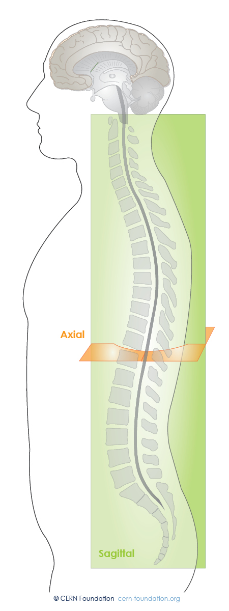
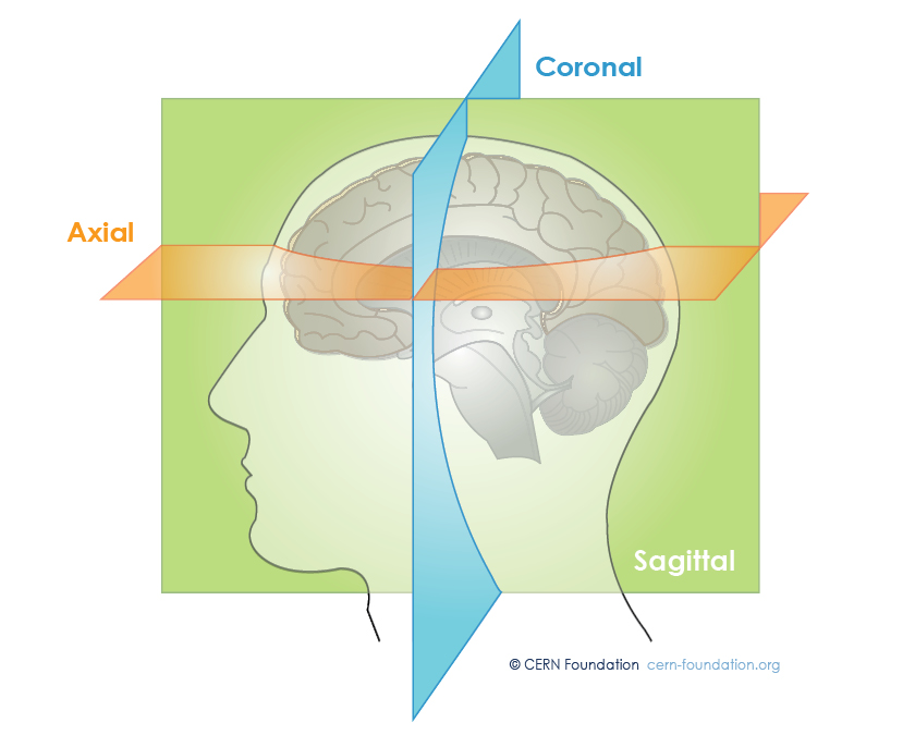
What types of images does an MRI take?
MRIs use three different views (see below) to show a tumor’s location and position. This helps your doctor compare the tumor with normal brain (or spinal cord) structures. Using these views, different scans can be done to produce exact images that best show the tumor and the area around the brain or spine. New techniques are being developed and tested on a regular basis.
The three types of MRI ‘views’ or images include:
- Sagittal: This is a vertical plane passing through the standing body from front to back
- Coronal: This is a vertical plane from head to foot that is parallel to the shoulders
- Axial: This is a straight line passing through a spherical body between two poles and that the body may revolve around
Other Tests
Sometimes a lumbar puncture (spinal tap) is necessary if there is concern that the tumor has spread into the cerebrospinal fluid.
Direct testing of the cerebrospinal fluid is done to look for the presence of tumor cells. In addition, an MRI of either the brain or spine may be ordered in addition to the baseline test to evaluate the entire CNS.

Coping with Scans
Hear from ependymoma survivor, Jennifer, as she shares how she copes with follow-up brain and spine scans and waiting for the results.
What causes ependymoma?
The ependymoma outcomes and risk project intends to answer the one question that plagues most all patients: Why did this happen to me?