Pediatric Ependymoma Images
What does an ependymoma look like in children?
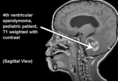
Sagittal image
Below is a pediatric ependymoma image – fourth ventricular ependymoma (T1 weighted with contrast):
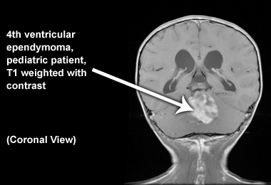
Coronal image
Below is an image of fourth ventricular ependymoma in a pediatric patient (T1 weighted with contrast):
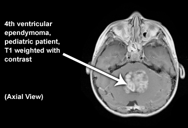
Axial image
Below is an image of fourth ventricular ependymoma in a pediatric patient (T1 weighted with contrast):
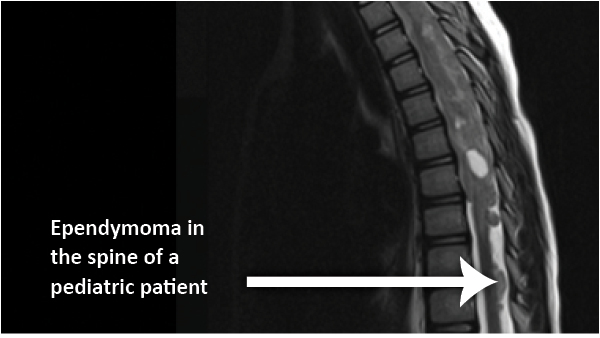
Spinal ependymoma
Below is an image of a spinal ependymoma in a pediatric patient: